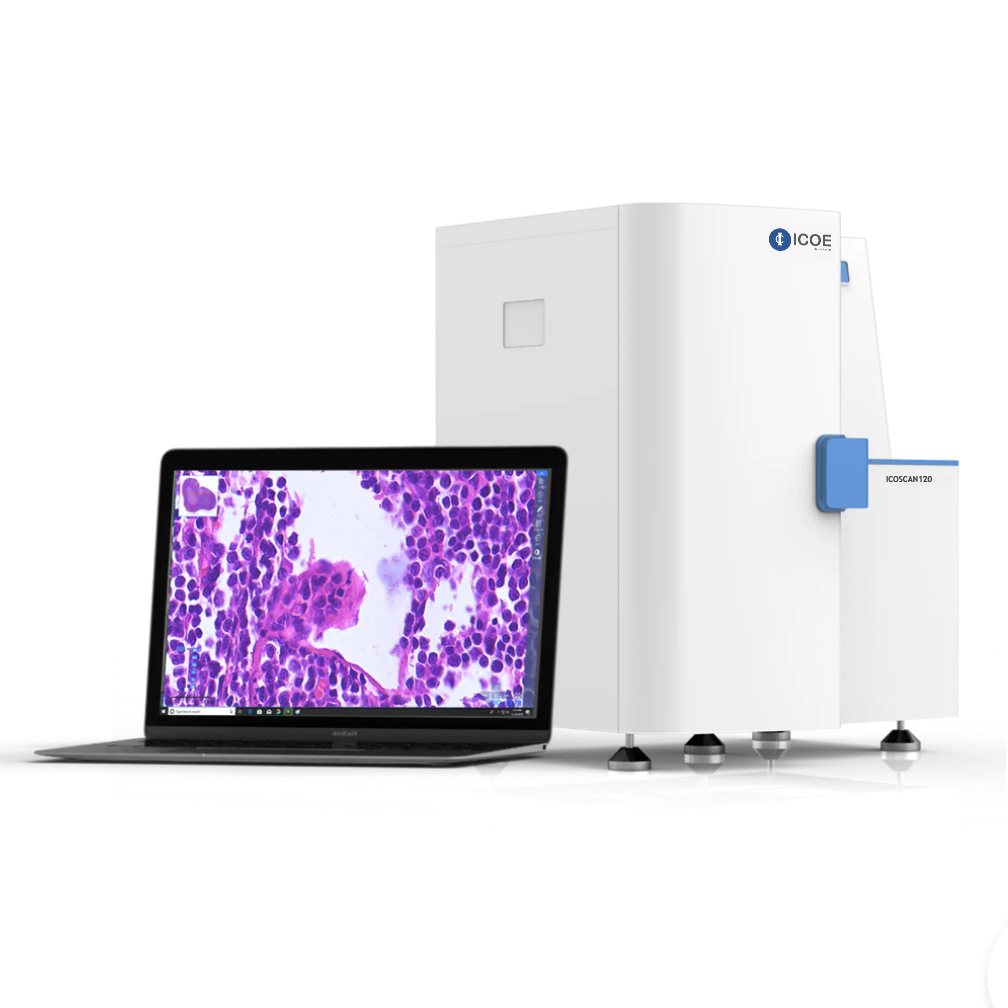
Description
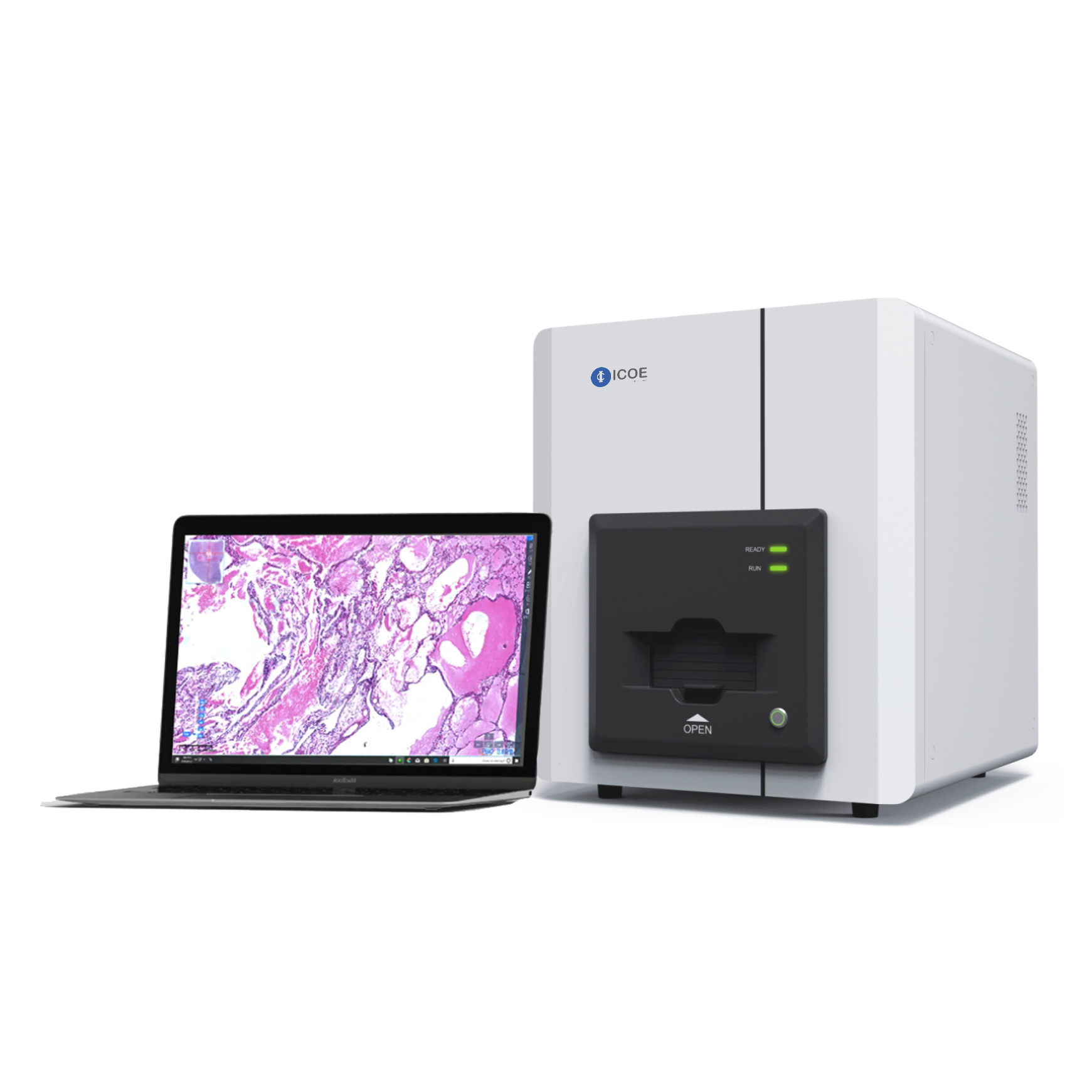
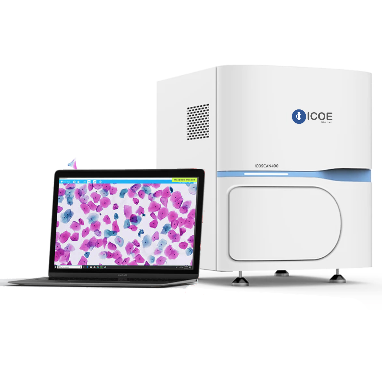
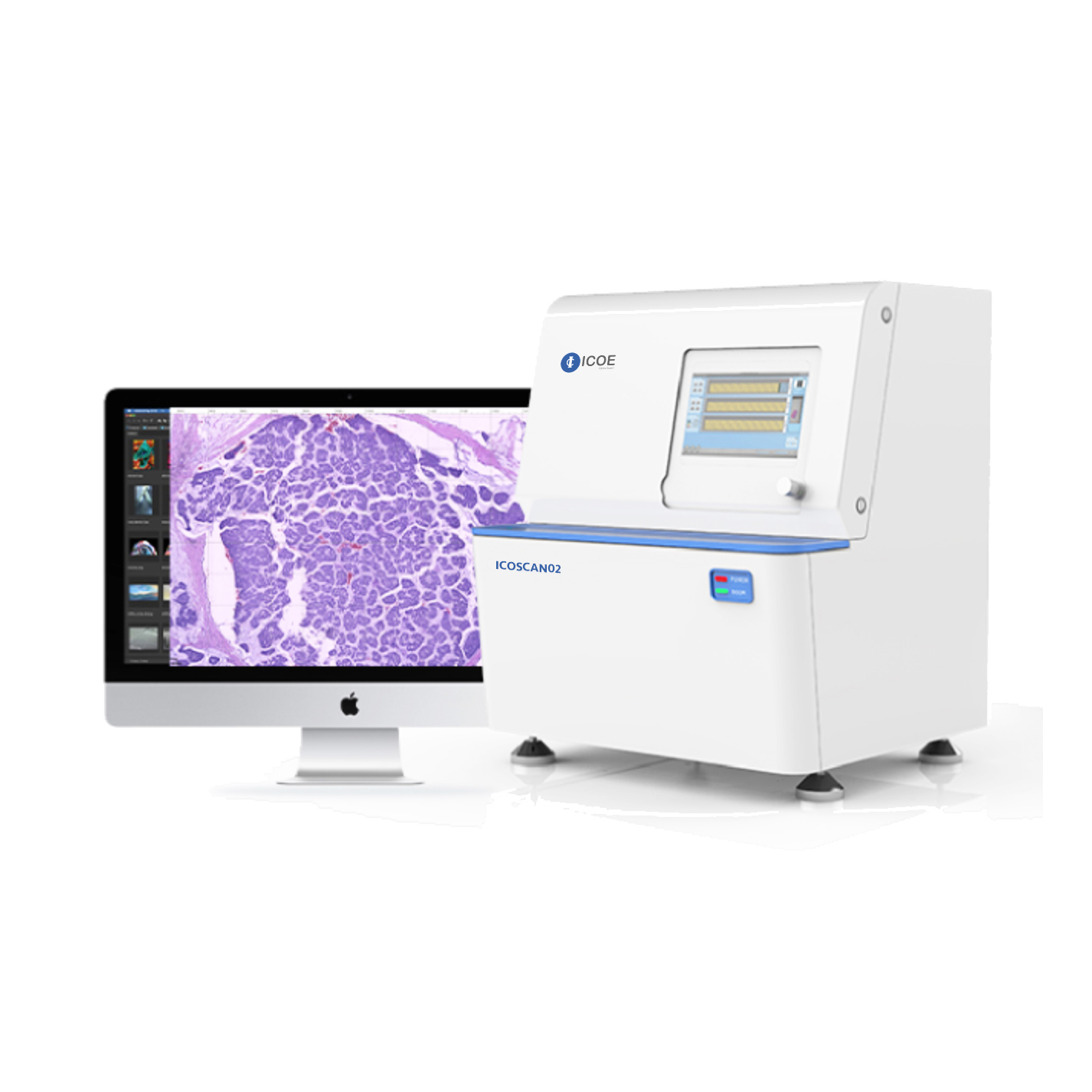
ICOE DMS-RPO digital pathological slice scanner is mainly composed of high-precision electromagnetic control system, optical imaging system, software control system and image management system. The fully automatic digital pathology slide scanner digitizes the slides, saves, browses, analyzes, and uploads them to the remote diagnosis platform through the image management system, enabling real-time remote pathological consultation.
Microscopic effect on screen – high-quality fully digital slides are the basis for accurate diagnosis.
The unique Rt-DFS (Realtime Dynamic Focus Scanning) technology ensures that the color of the image is truly restored, the image is clear, and the details are fully displayed.
Five slides Load Capacity, automatic identification of label information
Just take out the tray to load the slides, and place the barcode on the left side so that the device can automatically identify the code.
1. The startup of the instrument adopts the strong and weak electric segment control to ensure the safety of personnel and instruments
In order to prevent the “surge” of the 220V AC power from directly causing damage to the high-sensitivity electronic drive module and to ensure the safety of the staff’s power consumption, the strong and weak segmented control design is adopted.
2. Unique magnetic glass slide
The magnetic suction design ensures the precise positioning of the glass slide; avoids repeated friction during loading and unloading of the glass slide, ensures smooth loading and unloading, and prevents the slices from falling and breaking during loading and unloading.
3. High-speed and high-precision X/Y/2 three-dimensional stage system
3.1 Imported magnetic levitation linear motor: moving speed 3.2m/s, acceleration 8g.
3.2 High-precision full-closed-loop drive control system, X/Y-axis motion resolution is 100nm, and Z-axis motion resolution is 50nm.
4. Scanning software-specific features
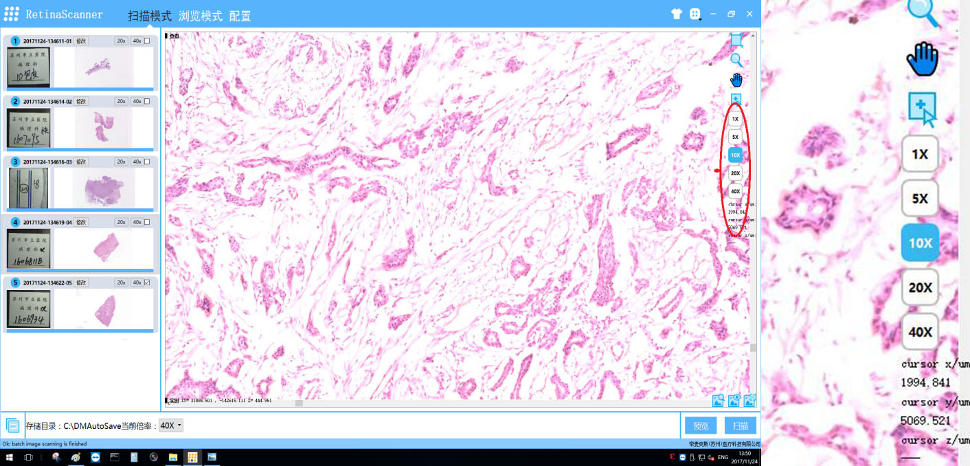
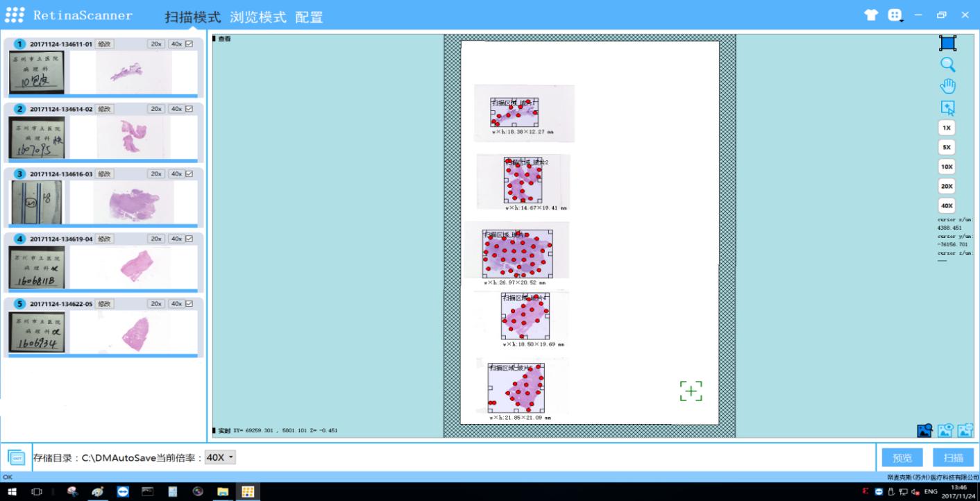
4.1 Scan and see: After the slice scan is completed, you can view the image immediately with the browsing function of the scanning software, and make a pathological diagnosis.
4.2 Real-time precise positioning and viewing of the slices that have just been scanned, which is equivalent to the microscope viewing function: the scanning software accurately positions the platform to move, and real-time positioning displays the slice images.
4.3 Real-time display of the current position parameters and tissue information parameters of the image.
4.4 When there are multiple pieces of tissue on the same slide, the software can intelligently identify the pieces, complete the scan independently, and fuse to generate a digital slice image.
5. Browse software
5.1 It has the function of slice library management: automatically detects slice files in the machine, and has the functions of retrieval, query and sorting.
5.2 Color distinguish slice browsing track: Different color marks locate the browsing track with different magnifications.
5.3 Mark ROI with different shapes, colors and line widths, and export the mark information as EXCEL files.
5.4 Area screenshots and whole screen screenshots of ≥300dpi can be taken to meet the publishing needs of journals and books; screenshots can be marked and exported.
DMS-PRO Digital Pathology Slide Scanner Parameter
| Model | DMS-PRO |
| Capacity | 5 slides |
| Scanning Mode | Area Scanning |
| Scanning Speed | <30s (20x, 15mmx15mm) |
| Scanning Magnifications | 20x/40x auto switch |
| Objective Lens | Plan apochromatic objective 20x, NA 0.75 |
| Scanning technology | Real-time Dynamic Focus Scanning |
| Resolution | 0. μm/pixel (20x); or 0.25 μm/pixel (40x) |
| Position Accuracy | X/Y: 100nm, Z: 50nm |
| Z-stack | ✓ |
| Image Format | JPG/TIFF/BMP |
| Movement Mechanism | Magnetic suspension drive, no noise, no abrasion |
| Auto focus | ✓ |
| Automatic identification of label information | ✓ |
| Automatic identification of sample area | ✓ |
| Multilayer scanning | ✓ |
| Cell morphometry | ✓ |
| Synchronized reading | ○ |
| Telepathology consultation | ○ |
| Incremental loading | ✓ |
| AI Recognition | ○ |
Note: “✓” means standard configuration, “○” means optional function, “-” without this function
Software Overview
Automatically and accurately identify tissue shape and tissue area, automatically skip blank areas, and automatically add pre-focus points to the identified tissue shape; it can perform intelligent block identification of multiple tissue blocks on the same slide and scan independently. The scanning area can be selected manually, the size of the area can be selected arbitrarily, and the size of the scanning area can be displayed in real time.
Support infinite zoom or fixed magnification zoom between full field of view magnification to 40×, seamless image, fast and smooth browsing, no lag, delay
