Description
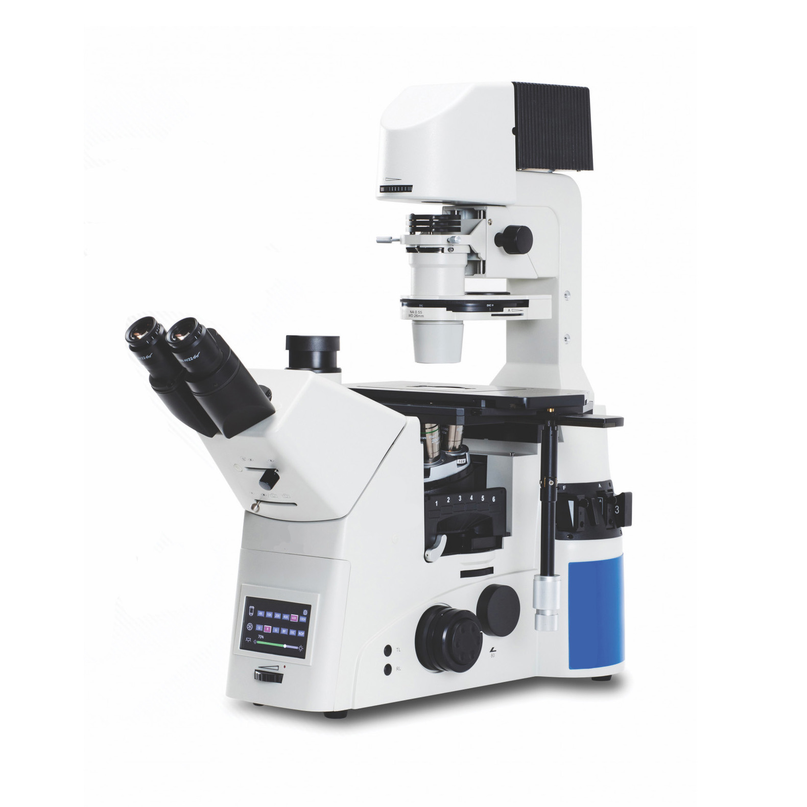
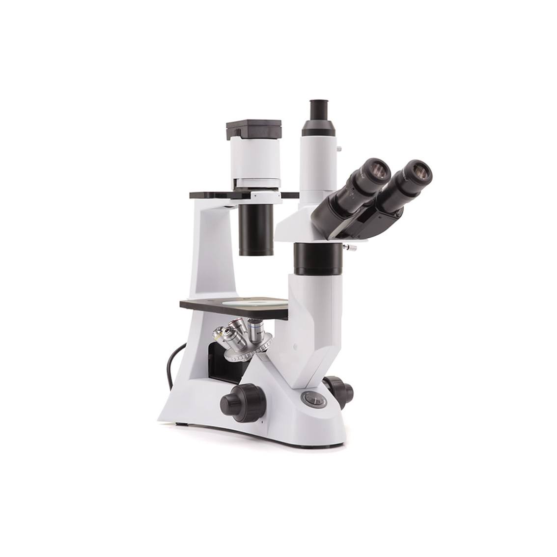
IB900 Series – ICOE’s flagship inverted microscope for life science research applications.
Empowering Discovery Advancing Results As a market leader in quality microscopy and imaging solutions, ICOE blends precision, performance and outstanding value like no other manufacturer can. We deliver superb yet affordable optical instrumentation and microscopy innovations that help advance the work, improve the outcomes and empower the achievements of the demanding professionals we are privileged to serve. No matter your industry, discipline or individual endeavor, count on ICOE microscopy, imaging solutions and accessories to exceed expectation as you achieve your goals. Market demands change and industries adapt, but our core principles never waver. More than any other provider. ICOE combines best-in-class optical performance with visionary innovation to engineer instrumentation of exacting design at exceptionally affordable price points. We produce products that inspire, empower and advance our evolving understanding of the world – in science, medicine, industry, research and education. Bringing next-generation solutions to the present to enable better outcomes, today and tomorrow.

Solution for your application

1). 6-way Nosepiece Intelligent sextuple nosepiece with click stops for precise positioning of the objective. Objective position is displayed on the LCD screen. A ballbearing track provides smooth movement
2). Phase Contrast High contrast method for viewing unstained cells in culture. Phase Contrast presents cells with a characteristic “halo” against a neutral background.
3). Filter Cube Turret The intelligent 6-position fluorescence filter cube turret allows quick and easy filter cube changing, and the filter cube position is indicated on the LCD screen.

4). LCD Screen Display The front-positioned, back-illuminated LCD screen displays objective, filter cube and light intensity setting
5). Light Selector Push-button switching from transmitted light to fluorescence reflected light.
6). Camera ports Top and side camera ports allow easy direct mounting of digital cameras.

7). Main camera port, on binocular head (same as above photo 6a)
8). Handle, for transportation
9). Handle, for transportation
10). Magnification changer, 1x-1.5x
11). Right side camera port (same as above photo 6b)
12). Universal condenser, for brightfield, PH and DIC
13). Viewing head, with standard WF10x/25mm eyepieces or WF10x/22mm
14). Slots for field diaphragm, aperture diaphragm, ND filters
15). Input port for fluorescence illuminator
16). Left side photo tube
17). Side photo tubes control (100/0; 20/80; 0/100)
18). Main photo tube control (100/0; 50/50; 0/100)
19). Bertrand lens insertion control
20). Main switch
21). Slot for analyzer
22). Focusing knobs
23). Three-layer mechanical stage
24). Transmitted illuminator brightness control

Multiple Observation Methods
Bright field Observation
Transmitted brightfield illumination is one of the most commonly used observation method in optical microscopy, and is ideal for fixed, stained specimens or other types of samples having high natural absorption of visible light. IB900 is fitted with high-efficiency LED brightfield illuminator, for the best outcome when using this technique.
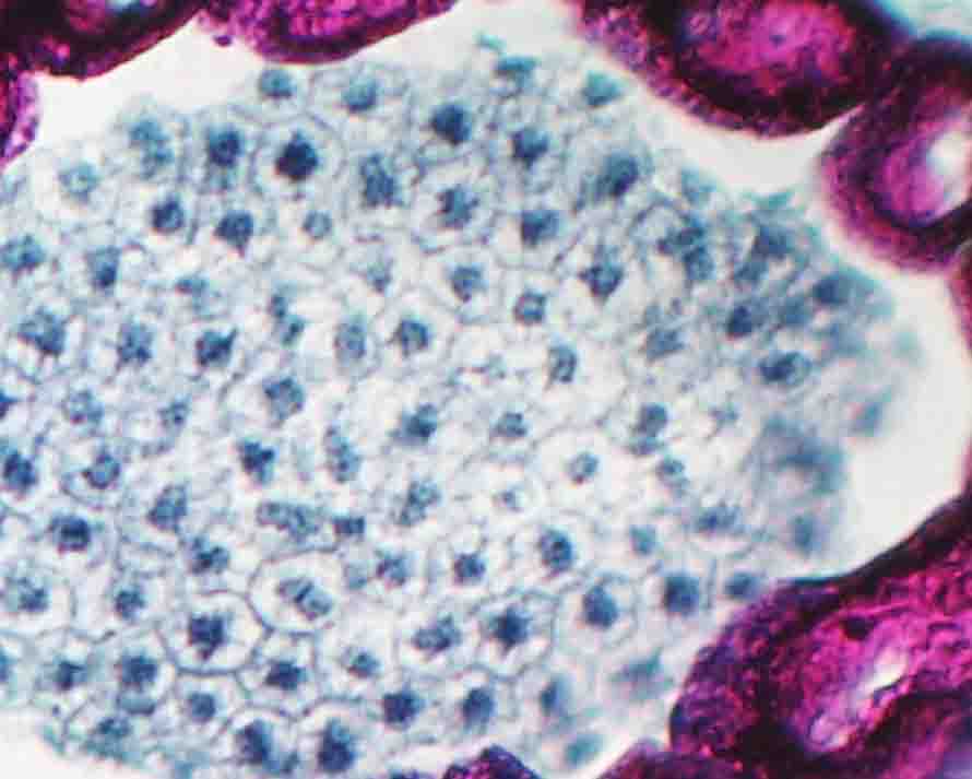
Capsella middle embry – Brightfield
Fluorescence Observation
The fluorescence microscopy is the most demanding technique in biology and biomedical sciences, as well as in material science. This method is capable to study organic and inorganic samples thanks to primary fluorescence (auto-fluorescence) or secondary (staining and labelling with fluorochromes). IB900 is tailored for application in research, clinical and pharmaceutical diagnostic field. Fluorescence illuminators available as mercury lamp.
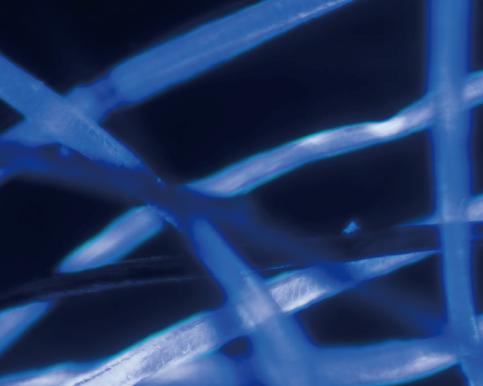
Cotton fibers – UV Fluorescence
DIC Observation
Differential Interference Contrast (DIC) is a microscopy technique that introduces contrast to images of specimens which have little or no contrast when observed using brightfield microscopy. The images produced using DIC have pseudo 3D-effect, making the technique ideal for many applications. DIC produces high resolution with good contrast. It is best for observing unstained samples.
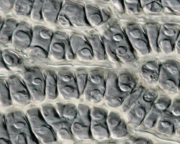
Sphagnum pores g- DIC
Phase Contrast Observation
Phase contrast microscopy is a particular technique applied in transparent, non-stainable, samples like culture of living cells, microorganisms, lithographic patterns, latex dispersions, fibers, asbestos and subcellular particles. It reveals many cellular structures that are not visible with simple brightfield microscope.
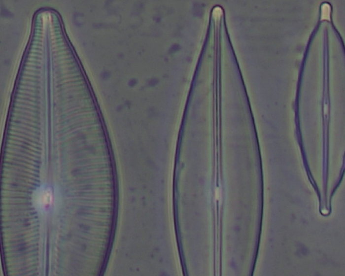
Diatoms – Phase contrast
Features
Revolving nosepiece for DIC and Fluorescence filter turret
The six-position nosepiece has a slot (in each of the six positions) for inserting DIC prisms. The filter turret can hold up to six fluorescence filter blocks. It is easily extractable, in order to facilitate the operation of inserting or replacing the filter blocks.
Condenser
Rotating N.A. 0.55 condenser with iris diaphragm. Working distance 26 mm. Supplied with phase contrast annuli for 10x/20x, 40x and 60x objectives and the DIC prisms for the 10x/20x/40x and 60x objectives. Three filter holders allow the insertion of Ø 38 mm filters in the optical path.
Illuminator arm
The arm of the transmitted illuminator is backward tilting up to 30 degrees and it allows to use flasks and big bottles.
Fluorescence equipment
A complete package of accessory dedicated to fluorescence technique is available as option
Mechanical stage and universal condenser
The wide 3-layer mechanical stage comes with several interchangeable plates for the use of Petri dishes, flasks and slides. The movement of the stage is controlled by a long tilting handle equipped with a pair of knobs for X/Y axes. The universal condenser is a 6-position type, designed for brightfield, phase contrast and DIC
Lens changer
A lens changer with a 1.5x lens allow intermediary magnifications of 1.5 times the standard objective magnifications. Switching from 20x to 30x or 40 to 60 and from 60x to 90x magnification can be done without the need to re-focus on the sample
Main photo tube and Bertrand lens
The main photo tube located on the binocular head is easily controllable by using its control knob. 3 positions selectable: 100/0, 50/50, 0/100. For phase contrast centering operations, a Bertrand lens is available and it can be easily inserted by means of a dedicated knob.
Optimize & automate experiments with motorized components
Upgraded your IB900 with software-controllable and motorized XYZ components from Prior, Ludl, Sutter or ASI. Ideal for live cell imaging applications.
Living Cell Imaging
The modularity and stability of the IB900 make it an ideal platform for live cell imaging. A variety of thirdparty accessories instantly transform the IB900 into an advanced multi-color fluorescence imaging solution. Choose from a selection of LED fluorescence illuminators, motorized XYZ components, filter wheels, antivibration tables, environmental chambers and cameras to perform techniques such as timelapse imaging. Z-stacks automatic scanning of well plates, stitching and more.

Specifications & Models
System Diagrams
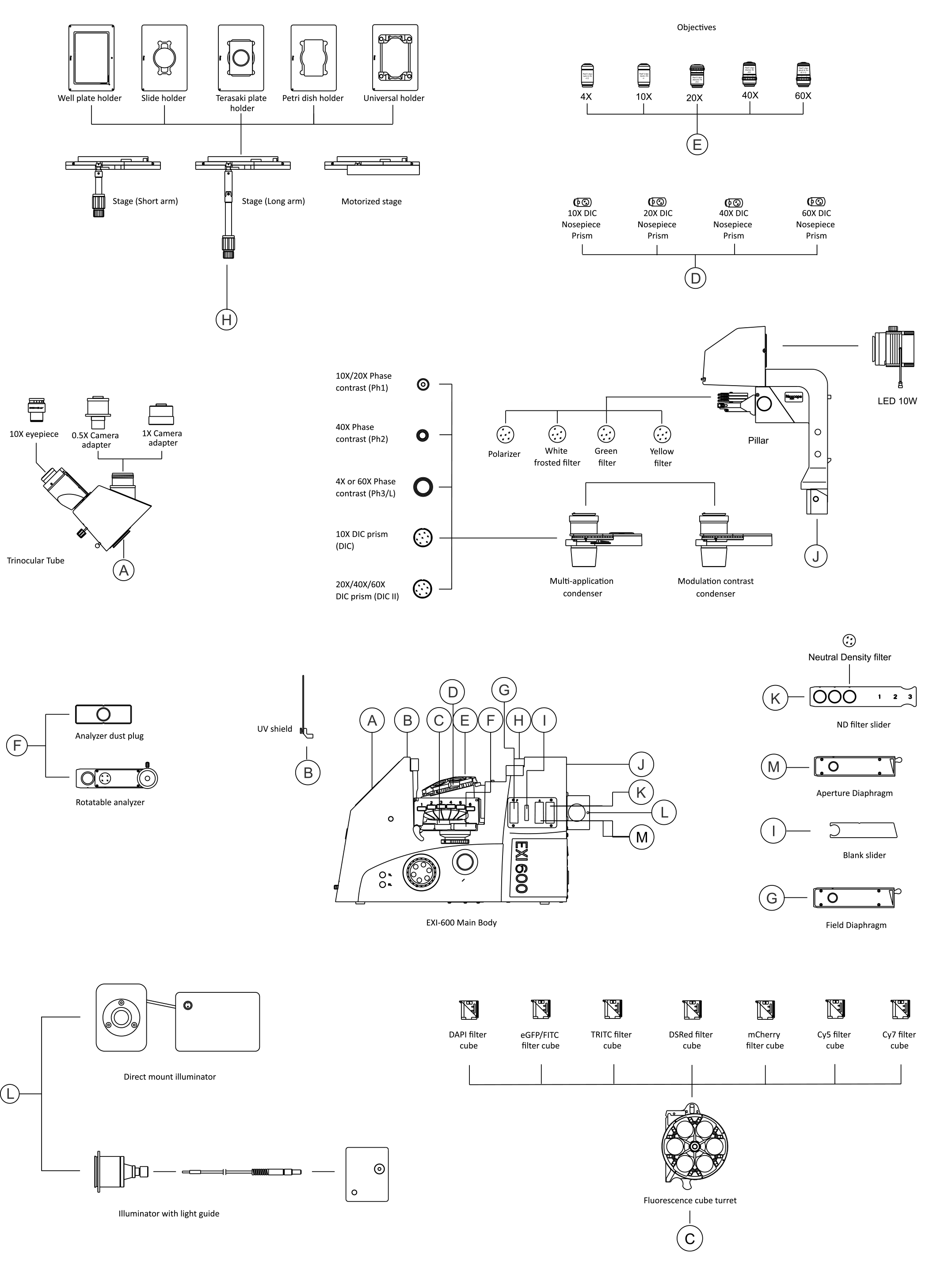
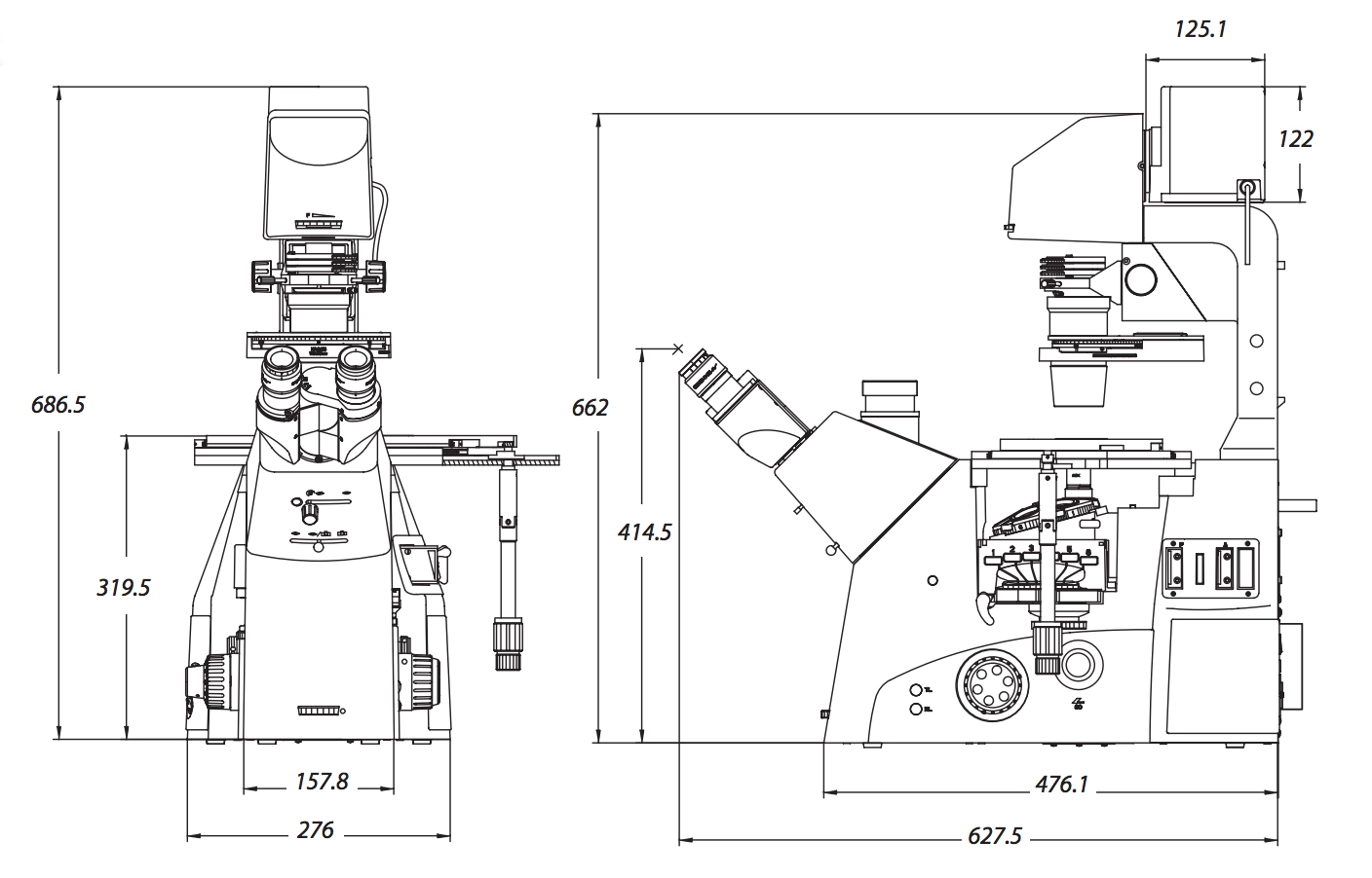
Product Dimension
