Description
Introduction
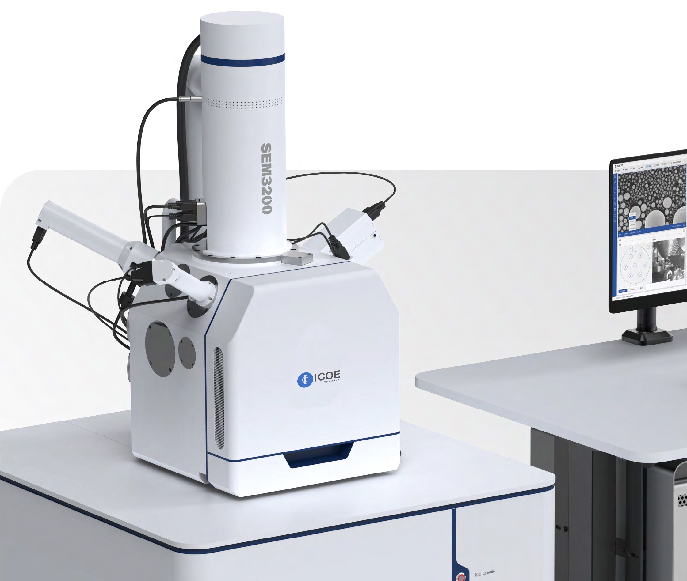
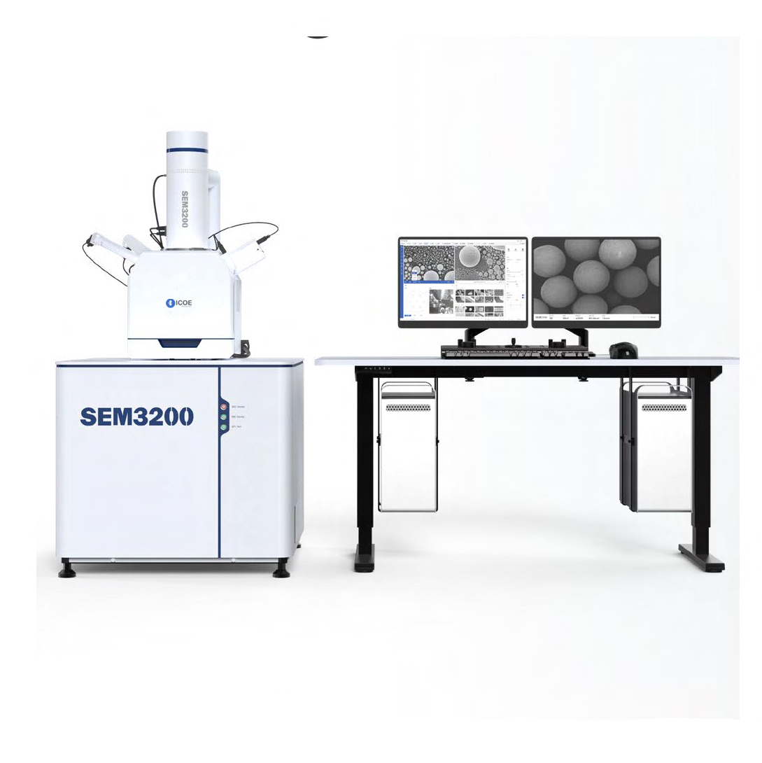
SEM3200 is a tungsten filament scanning electron microscope with high performance and wide application. It has excellent imaging quality capabilities in both high and low vacuum modes. It also has a large depth of field with a user‑friendly environment to characterize samples.What’s more, rich scalability helps the users to explore the world of microscopic imaging.

Note: * is optional item
Product Advantages
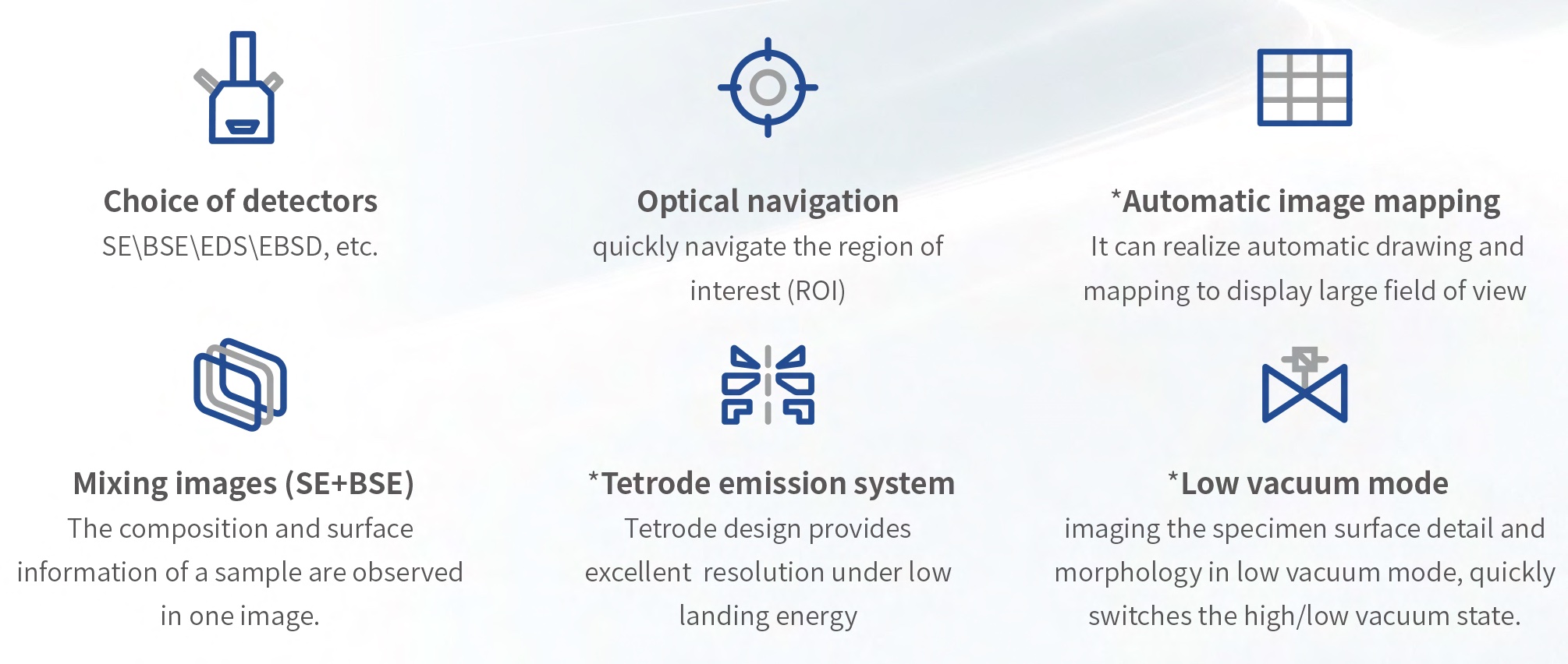
Note: * is optional item
Product Features
> Low Voltage
Imaging of the carbon material specimen, the penetration depth of the specimen is small under lo w voltage(3kV) of secondary electron mode, the more surface de tail information.

> Low Vacuum
The filer tube material has poor electrical conductivity, and charging with out coating in high vacuum condition, but can be directly imaged in low vacuum condition.

> Large Field of View
With a large field of vie w, the overall morphology and head structure de tails of ladybug c an be easily obtained, and the cross-scale analysis can be displayed.

> Optical Navigation
See where you want, the navigation is easier to achieve. The standard configuration in-chamber camera can take high-definition photos of the sample stage,and samples can be quickly located.
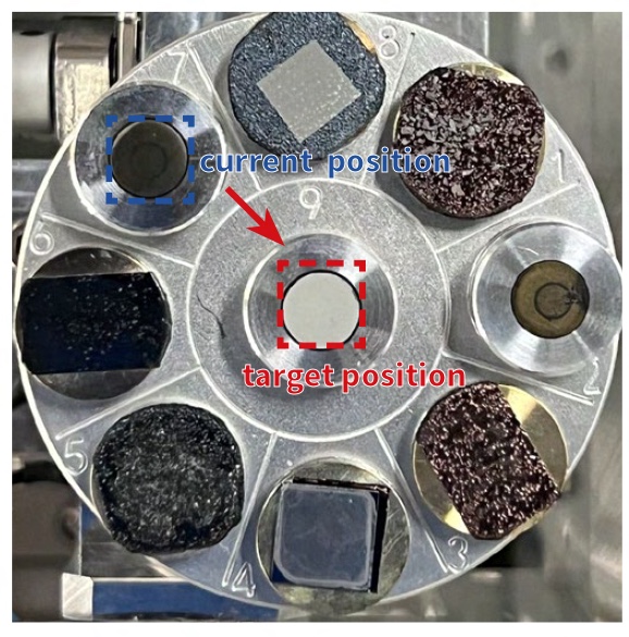
> Collision Prevention Technology
Multi-dimensional anti-collision scheme:
1.The height of the sample can be input ted manually, and the distance between the sample top and the end of the objective lens can be accurately controlled to prevent collision.
2. Based on image recognition and dynamic capture technology, you can acquire the real-time image in chamber during the movement of the sample.
3.*Hardware anti-collision, which can stop the motor immediately at the moment of collision and reduce collision damage.
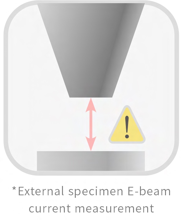
Note: * is optional
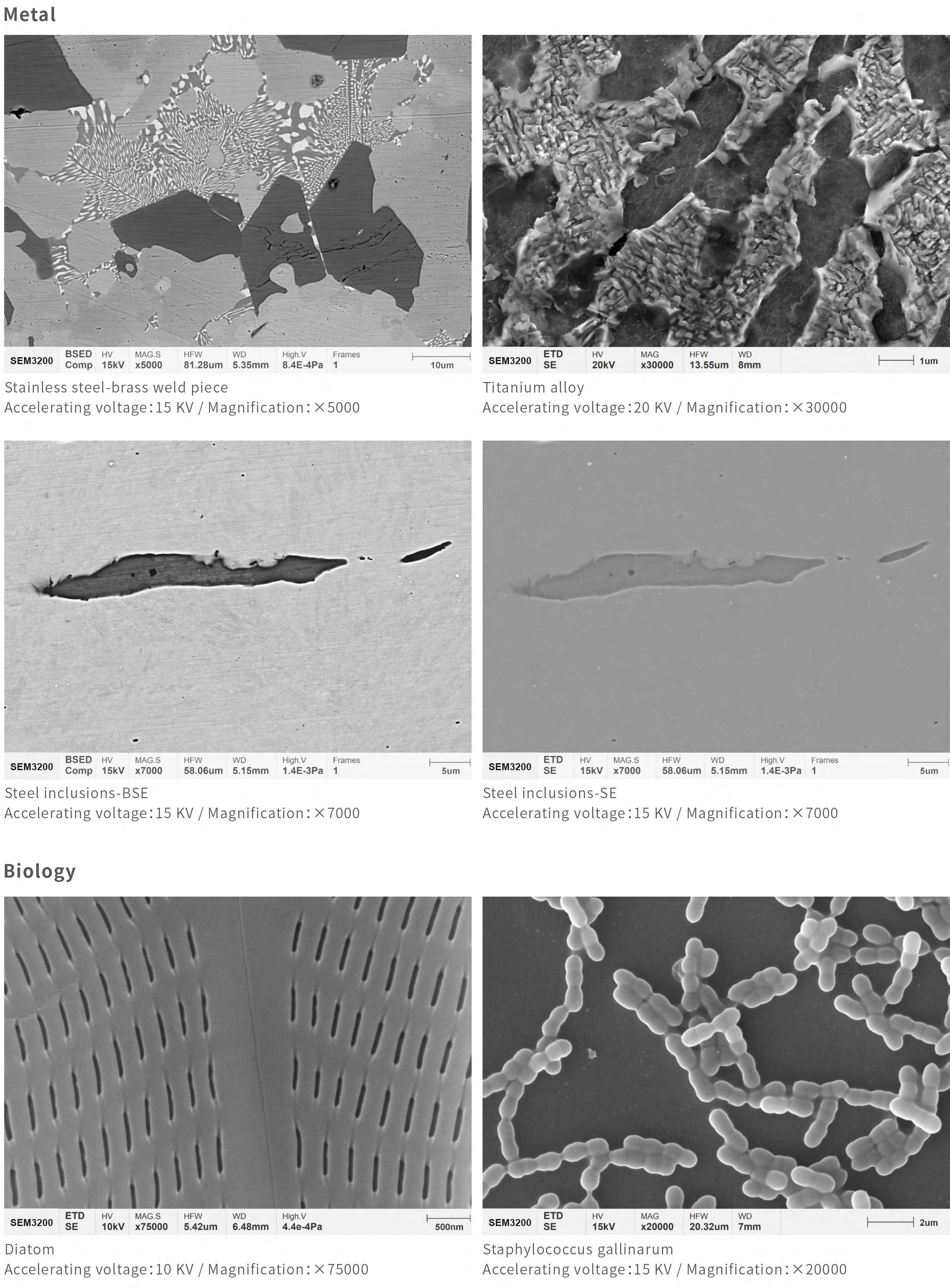
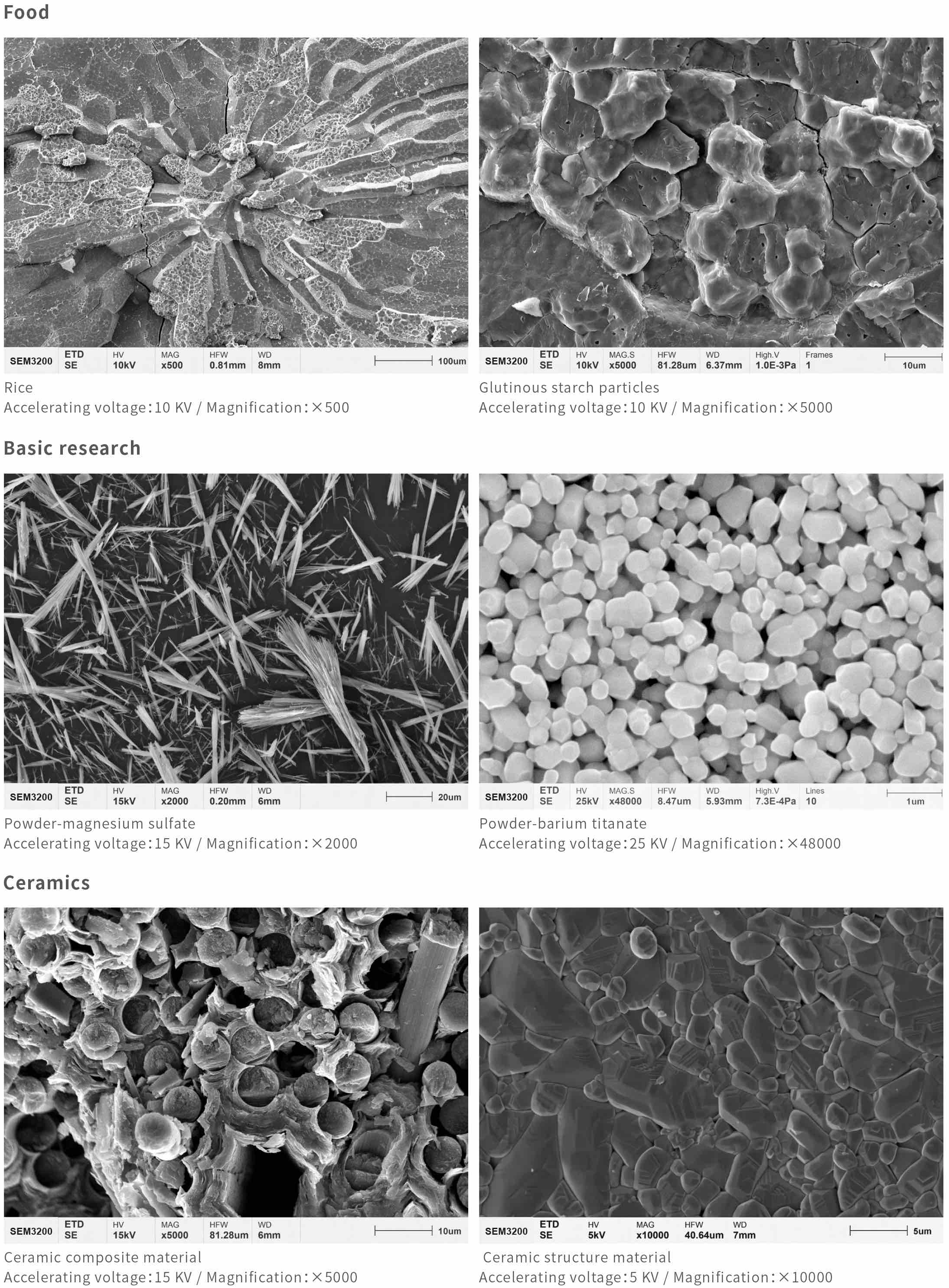
Application Field & Cases
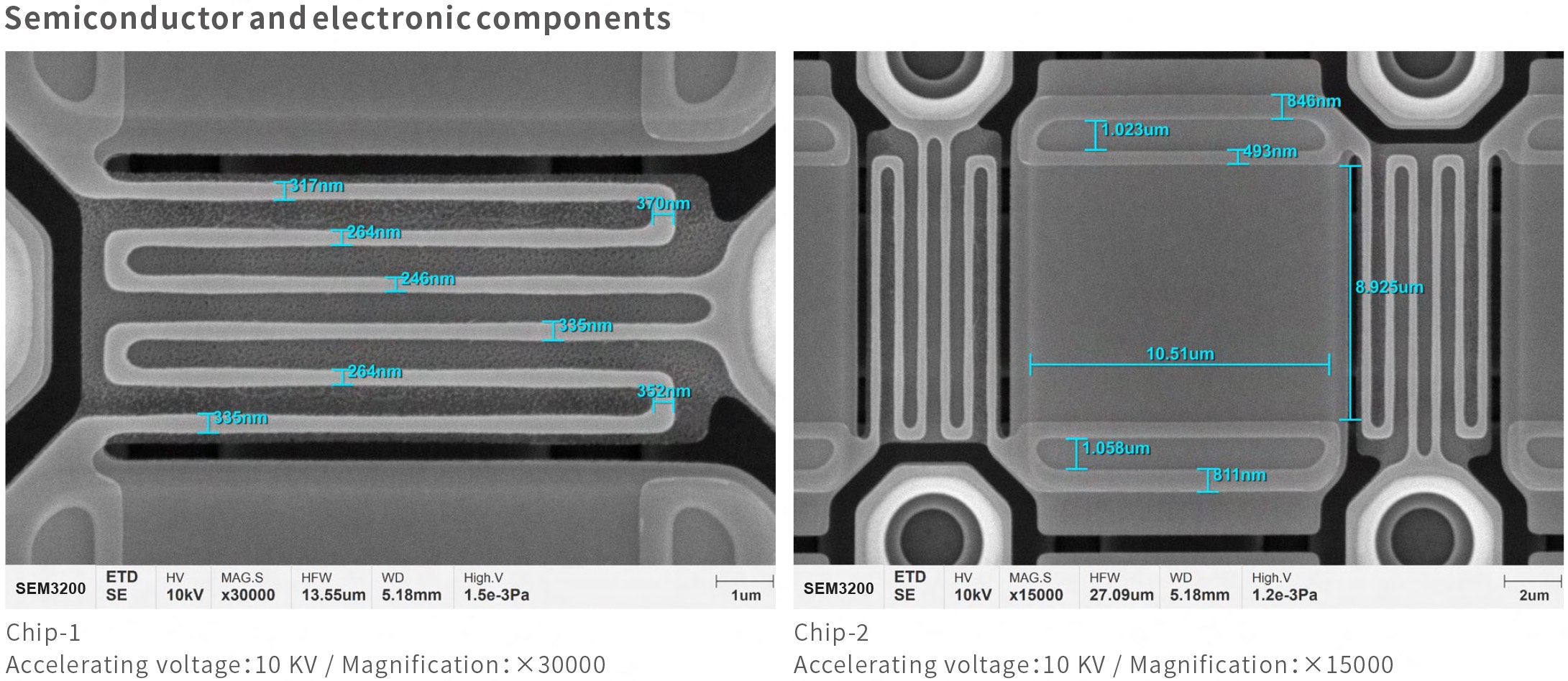
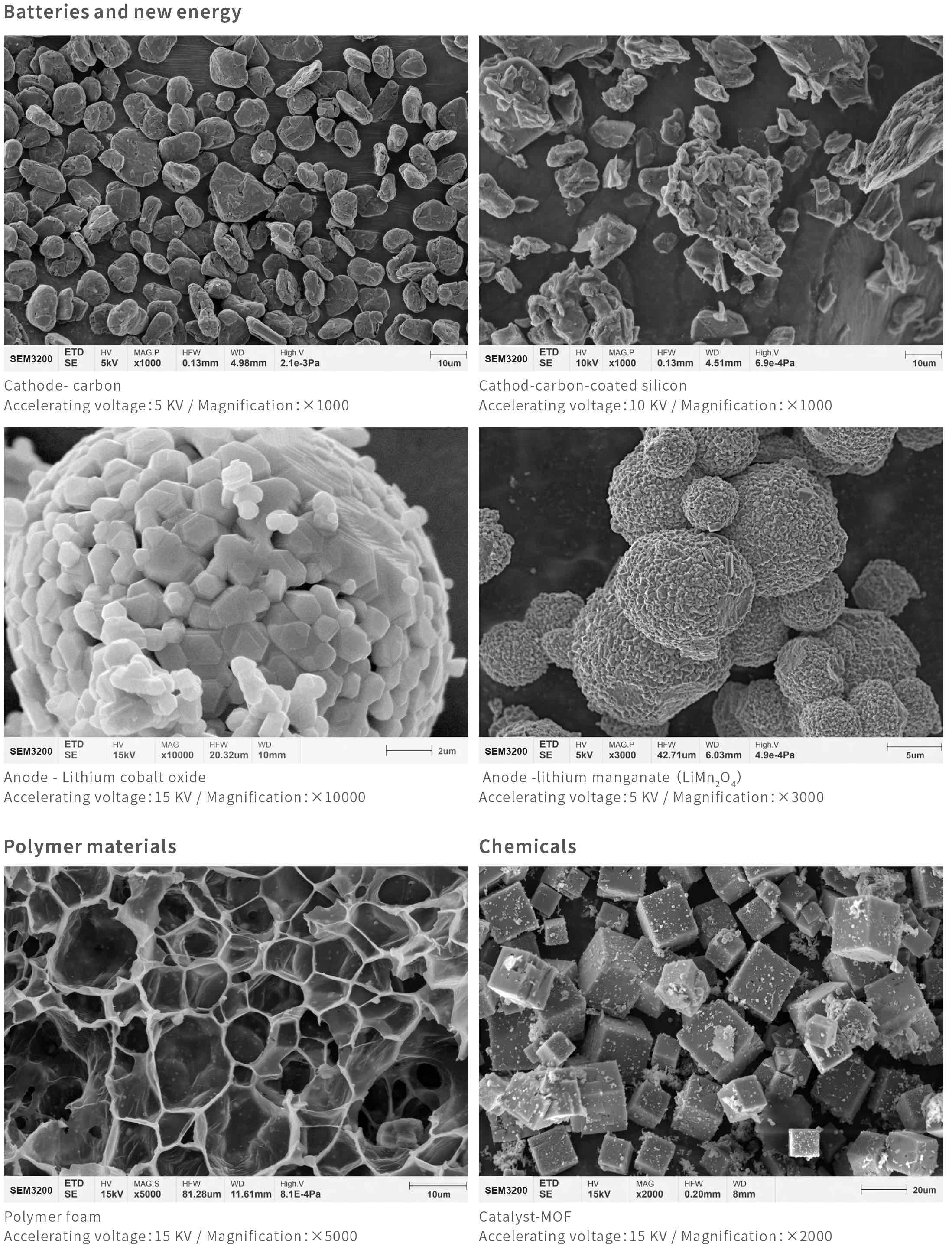
Specifications
| Models | SEM3200A | SEM3200 | ||
| Electro-Optical Systems | Electron Gun | Pre-aligned medium-sized fork-type tungsten filament | ||
| Resolution | High Vaccum | 3 nm @ 30 kV (SE) | ||
| 4 nm @ 30 kV (BSE) | ||||
| 8 nm @ 3 kV (SE) | ||||
| *Low Vaccum | 3 nm @ 30 kV (SE) | |||
| Magnification | 1-300,000x (Film Magnification) | |||
| 1-1000,000x (Screen Magnification) | ||||
| Acceleration Voltage | 0.2 kV ~ 30 kV | |||
| Imaging Systems | Detector | Secondary Electron Detector (ETD) | ||
| Backscattered electron detector (BSED), low vacuum secondary electron detector, *energy spectrometer EDS, etc. | ||||
| Image Format | TIFF, JPG, PNG | |||
| Vacuum System | Vacuum Model | High Vacuum | Better than 5×10⁻⁴ Pa | |
| Low Vacuum | 5 ~ 1000 Pa | |||
| Control Mode | Automatic Valve | |||
| Turbomolecular Pump | ≥ 240 L/S | |||
| Mechanical Pump | 200 L/min (50 Hz) | |||
| Sample Chamber | Camera | Optical Navigation | ||
| Monitoring in the Sample Chamber | ||||
| Sample Stage Configuration | Three Axis Automatic | Five Axis Automatic | ||
| Distance | X: 120 mm | X: 120 mm | ||
| Y: 115 mm | Y: 115 mm | |||
| Z: 50 mm | Z: 50 mm | |||
| / | R: 360° | |||
| / | T: -10° ~ +90° | |||
| Software | Operating System | Windows | ||
| Navigations | Optical Navigation, Gesture Quick Navigation | |||
| Automatic Functions | Auto Brightness Contrast, Auto Focus, Automatic Dissipation | |||
| Special Functions | Intelligent Assisted Dispersion, *Large-Scale Image Stitching (Optional accessories) | |||
| Installation Requirements | Space | L≥ 3000 mm, W ≥ 4000 mm, H ≥ 2300 mm | ||
| Temperature | 20°C (68°F) ~ 25°C (77°F) | |||
| Humidity | ≤ 50 % | |||
| Power Supply | AC 220 V(±10 %), 50 Hz, 2 kVA | |||
Note: * is optional item

